 Image credit: ISCB
Abstract
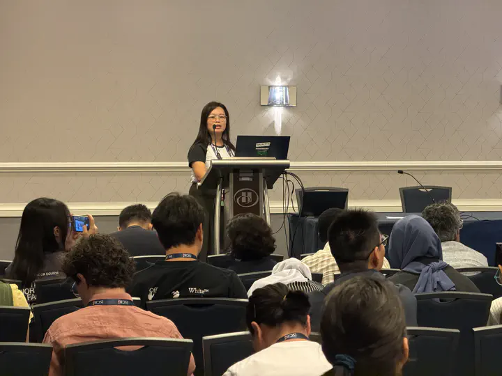
Cellular phenotypes in the tumor–immune microenvironment are central to tumor progression and response to immune checkpoint inhibitors, yet they are largely shaped by spatial tumor–immune colocalization. Spatial profiling technologies such as CODEX and PhenoCycler Fusion can resolve high-dimensional cell states, but remain limited by cost and throughput. In contrast, haematoxylin and eosin (H&E) histology is widely available but lacks direct phenotypic labels. Building on FineST, Hist2Pheno infers nuclei-resolved cellular phenotypes from routine histological images, enabling high-resolution spatial analysis without matched molecular profiling. This 20-minute talk was given in the Computational Systems Immunology (CSI) COSI session on Spatial Transcriptomics & Imaging (12:20–12:40, 13 July 2026) at the 34th ISMB conference.
Talk. Hist2Pheno enables nuclei-resolved phenotypic prediction from histological images in esophageal squamous cell carcinoma.
Session. Computational Systems Immunology (CSI) COSI — Spatial Transcriptomics & Imaging, 12:20–12:40, 13 July 2026, Washington Hilton.
Code. Hist2Pheno

Lingyu Li
Postdoctoral Fellow
Research in bioinformatics, including spatial transcriptomics, sparse statistical learning, and biomarker discovery.